Stereotactic Biopsy Brain Tumor
A single MRI scan can change everything.
One moment, life is moving normally. Next, a doctor points to a tiny abnormal area on a brain scan and says, “We need to investigate this further.”
For most patients and families, this is where uncertainty begins. Is it a benign tumor? Is it cancerous? Could it be an infection? Is surgery necessary? What happens next?
Modern MRI and CT scans are remarkably advanced, but even the most detailed images cannot always provide a definitive diagnosis. Different brain conditions can appear surprisingly similar on imaging studies. A lesion that looks like a tumor
may actually be inflammation. A suspected cancer may turn out to be a treatable infection. Likewise, two tumors that appear nearly identical on scans can behave very differently and require completely different treatment approaches.
This is why obtaining an accurate diagnosis before starting treatment is one of the most important principles in modern neuro-oncology. Rather than making assumptions based solely on imaging findings, specialists often rely on stereotactic biopsy—a highly precise, minimally invasive procedure that allows them to obtain tissue samples from deep within the brain.
Today, Stereotactic Biopsy Brain Tumor in Mumbai has become an essential diagnostic tool, helping neurosurgeons determine exactly what they are treating before recommending surgery, radiation therapy, chemotherapy, or other advanced interventions.
Why Modern Brain Tumor Care Starts With Certainty?
Imagine being prescribed treatment without knowing the exact disease.
That approach would never be acceptable in modern medicine.
Brain tumors vary significantly in:
- Growth rate
- Aggressiveness
- Treatment response
- Long-term prognosis
- Surgical complexity
Even tumors arising from the same area of the brain can require completely different management plans.
A tissue diagnosis helps doctors understand:
- The exact tumor type
- Whether the lesion is benign or malignant
- How aggressive the tumor is
- Whether molecular markers are present
- Which treatment strategy offers the best chance of success
Without a biopsy, important treatment decisions may be based on incomplete information.
What Is a Stereotactic Brain Biopsy?
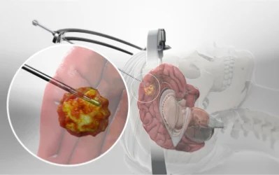
A stereotactic brain biopsy is a sophisticated neurosurgical procedure designed to obtain a small tissue sample from an abnormal brain lesion using advanced imaging and computer-guided navigation.
Unlike traditional open brain surgery, the objective is not to remove the tumor immediately. Instead, the goal is to obtain enough tissue for detailed pathological examination.
The procedure uses:
- MRI imaging
- CT imaging
- Three-dimensional computer mapping
- Real-time navigation systems
- Precision biopsy instruments
These technologies work together to guide the surgeon to the exact target location while minimizing disruption to surrounding brain tissue.
Because of its accuracy, stereotactic biopsy has become one of the most trusted methods for diagnosing complex brain lesions.
Which Patients May Benefit From a Brain Biopsy?
Not every brain tumor requires immediate surgical removal.
In many situations, obtaining a diagnosis first is the safest and most logical approach.
A stereotactic biopsy may be recommended when:
The Lesion Is Deep Inside the Brain
Certain tumors are located in regions that are difficult to access through conventional surgery.
The Diagnosis Is Unclear
Imaging studies may suggest multiple possible conditions.
The Tumor Is Close to Critical Brain Structures
Areas responsible for speech, movement, memory, or vision require careful planning before major intervention.
Multiple Lesions Are Present
Biopsy can help determine whether lesions represent metastatic disease, infection, lymphoma, or another condition.
Treatment Decisions Depend on Pathology
The biopsy findings often determine whether surgery, radiation, chemotherapy, or observation is most appropriate.
The Technology Behind Pinpoint Accuracy
One of the most remarkable aspects of stereotactic biopsy is its precision.
Before the procedure begins, advanced MRI and CT scans create a three-dimensional map of the patient’s brain.
Using specialized software, neurosurgeons can:
- Identify the safest pathway
- Avoid critical neurological structures
- Target the most informative part of the lesion
- Reduce procedural risks
The navigation system functions much like a GPS, guiding the surgeon with extraordinary accuracy.
This technology has significantly improved the safety and reliability of Brain tumor biopsy Mumbai procedures.
What Happens During the Procedure?
Many patients are surprised to learn that stereotactic biopsy is far less invasive than traditional brain surgery.
Detailed Preoperative Planning
High-resolution imaging studies are reviewed and integrated into the navigation system.
Precise Target Mapping
The exact biopsy location is selected based on imaging characteristics.
Small Surgical Opening
A tiny incision and a small opening in the skull are created.
Tissue Collection
A specialized biopsy needle is guided directly to the target area.
Several tissue samples may be collected to improve diagnostic accuracy.
Pathological Examination
The samples are sent to specialized laboratories where experts analyze the tissue under a microscope and perform advanced testing if required.
The entire process is designed to obtain critical diagnostic information while minimizing surgical trauma.
Why Tissue Analysis Matters?
The tissue obtained during biopsy provides far more information than a simple yes-or-no diagnosis.
Modern pathology can reveal:
Tumor Classification
Determining the exact tumor type.
Tumor Grade
Understanding how aggressively the tumor is likely to behave.
Molecular Characteristics
Identifying genetic markers that may influence treatment decisions.
Treatment Sensitivity
Predicting which therapies are likely to be most effective.
This information forms the foundation of personalized brain tumor care.
Recovery Is Usually Faster Than Patients Expect
One of the reasons stereotactic biopsy is widely used is its minimally invasive nature.
Compared to open brain surgery, patients often experience:
- Smaller incisions
- Less discomfort
- Shorter hospital stays
- Faster recovery
- Reduced disruption to daily life
Most individuals are able to resume routine activities relatively quickly, although recovery timelines vary based on individual circumstances.
Follow-up appointments ensure proper healing and discussion of pathology results.
From Diagnosis to Personalized Treatment
The biopsy result serves as a roadmap for future treatment.
Depending on the findings, the next step may involve:
Observation
Some slow-growing lesions may only require monitoring.
Brain Tumor Surgery
Certain tumors may be suitable for surgical removal.
Radiation Therapy
Often used for selected benign and malignant tumors.
Chemotherapy
Recommended for specific cancer types.
CyberKnife Radiosurgery
A highly precise, non-invasive treatment option for carefully selected cases.
The goal is always to provide treatment tailored to the patient’s specific diagnosis rather than relying solely on imaging appearances.
Why Patients Trust Dr Manish Baldia?
Accurate diagnosis is the cornerstone of successful brain tumor treatment, and achieving that diagnosis requires both advanced technology and expert clinical judgement.
Dr Manish Baldia is a neurosurgeon and functional neurosurgery specialist with 11 years of experience in treating complex neurological conditions. After completing his MBBS and MCh in Neurosurgery, he developed expertise in advanced neurosurgical procedures that prioritize precision, safety, and individualized patient care.
His clinical interests include stereotactic neurosurgery, minimally invasive brain tumor surgery, CyberKnife radiosurgery, Deep Brain Stimulation (DBS), spinal cord tumor surgery, and functional neurosurgical procedures. Practicing across Mumbai, Pune, Nagpur, and Hyderabad, he has helped numerous patients navigate challenging neurological diagnoses through evidence-based treatment strategies.
In addition to his clinical work, Dr Manish Baldia is the innovator behind cost-effective cranioplasty implant solutions through NaisBrain Pvt. Ltd., reflecting his commitment to making advanced neurosurgical care more accessible. For patients seeking Stereotactic Biopsy Brain Tumor in Mumbai, his focus remains on obtaining accurate diagnoses through the safest and most effective diagnostic approaches available today.
When a brain lesion is discovered, the most important question is often the simplest one: “What exactly are we dealing with?” The answer can shape every treatment decision that follows. Through Stereotactic Biopsy Brain Tumor in Mumbai, neurosurgeons can obtain crucial diagnostic information with remarkable precision while minimizing disruption to healthy brain tissue. By transforming uncertainty into clarity, this advanced procedure allows patients and doctors to move forward with confidence, knowing that future treatment decisions are based on accurate scientific evidence. With extensive experience in stereotactic neurosurgery and brain tumor management, Dr Manish Baldia helps patients take the first and often most important step toward effective neurological care.
The Most Frequently Asked Questions
No. A stereotactic biopsy is a minimally invasive diagnostic procedure designed to obtain tissue samples rather than remove the entire tumor.
Although MRI provides valuable information, many lesions appear similar on scans. Tissue examination is often required for a definitive diagnosis.
Modern stereotactic biopsy procedures are considered highly safe when performed by experienced neurosurgical teams using advanced navigation systems.
The duration varies depending on the lesion and surgical plan, but most procedures are completed within a few hours.
Preliminary results are often available within a few days, while specialized testing may take longer.


